What Makes an Action Potential Rise to +35 Mv? What Makes It Drop Again After This Peak?
half-dozen Action Potentials
As covered in Chapter ane, the activeness potential is a very brief change in the electric potential, which is the difference in charge between the inside and outside of the cell. During the action potential, the electrical potential across the membrane moves from a negative resting value to a positive value and back.
Propagation
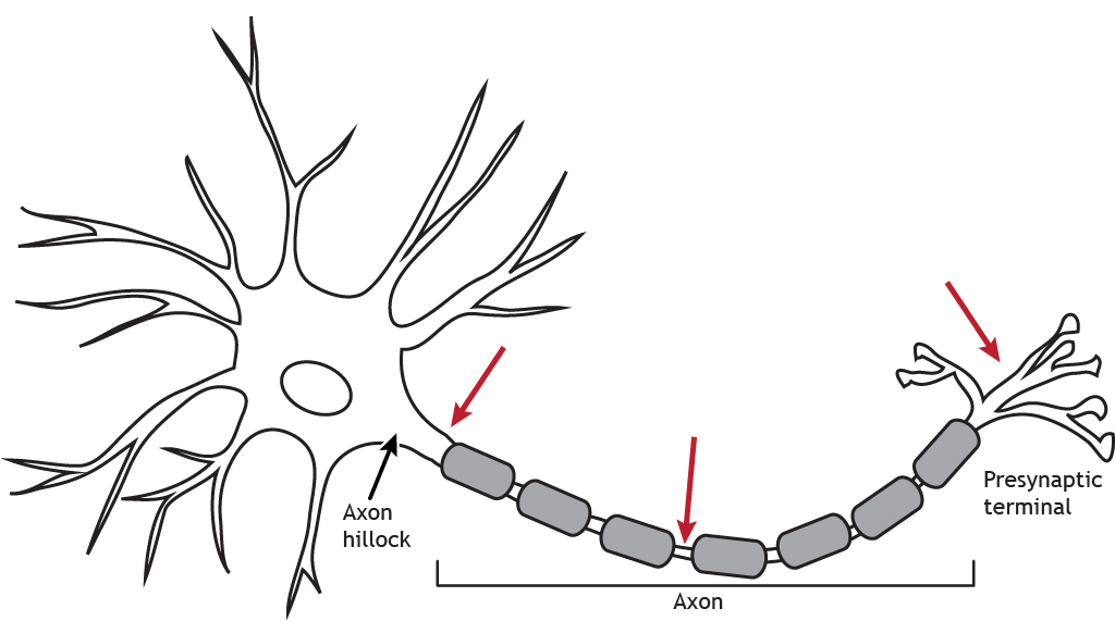
The propagation of the action potential from the axon hillock down the axon and to the presynaptic terminal results in release of chemical neurotransmitters that communicate with a postsynaptic neuron.
Blitheness 6.1. The activeness potential moves down the axon beginning at the axon hillock. The action potential moving down a myelinated axon will jump from one Node of Ranvier to the next. This saltatory conduction leads to faster propagation speeds than when no myelin in present. When the action potential reaches the synaptic concluding, it causes the release of chemical neurotransmitter. 'Activeness Potential Propagation' by Casey Henley is licensed under a Creative Commons Attribution Non-Commercial Share-Akin (CC-BY-NC-SA) 4.0 International License. View static image of animation.
Voltage-Gated Ion Channels
The change in membrane potential during the action potential is a function of ion channels in the membrane. In the previous lessons, we take learned nigh the principles of ion movement and have discussed non-gated (leak) channels at rest, likewise as ion channels involved in the generation of postsynaptic potentials. In this affiliate, we will examine a different type of ion channel: voltage-gated ion channels. For our purposes, these channels are located primarily at the axon hillock, forth the axon and at the terminal. They are necessary for the propagation of the action potential.
Voltage-gated channels allow ions to cross the membrane using the same ion movement principles covered in previous lessons. The master difference between voltage-gated channels and leak channels are how they are opened or "gated". Voltage-gated channels open when the cell'due south membrane potential reaches a specific value, chosen threshold. The neuron reaches threshold after enough EPSPs summate together.
Animation six.2. As EPSPs calculate, a result of ion movement not shown in the animation, the cell's membrane potential will depolarize. Reaching threshold causes voltage-gated ion channels to open. One time the channels are open up, ions will movement toward equilibrium. In the animation, sodium ions catamenia inward. The dotted, blue channels represent voltage-gated sodium channels; the striped, greenish channels stand for voltage-gated potassium channels; the solid yellow channels represent chloride channels. 'Voltage-Gated Aqueduct' by Casey Henley is licensed under a Creative Commons Attribution Non-Commercial Share-Alike (CC-BY-NC-SA) 4.0 International License. View static image of blitheness.
The Activeness Potential
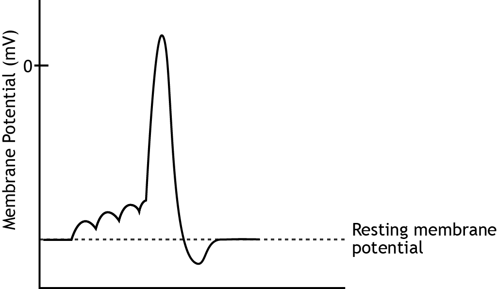
The action potential begins when the cell's membrane potential reaches threshold. In one case initiated in a healthy, unmanipulated neuron, the action potential has a consistent structure and is an all-or-nothing upshot. Information technology will run through all the phases to completion.
The rising phase is a rapid depolarization followed past the overshoot, when the membrane potential becomes positive. The falling phase is a rapid repolarization followed past the undershoot, when the membrane potential hyperpolarizes past rest. Finally, the membrane potential will return to the resting membrane potential.

Rising Phase
The rise phase is caused past the opening of voltage-gated sodium channels. These ion channels are activated once the prison cell'due south membrane potential reaches threshold and open immediately. The electrochemical gradients drive sodium into the jail cell causing the depolarization.
Animation 6.three. Voltage-gated sodium channels open once the jail cell's membrane potential reaches threshold. The rapid influx of sodium results in a large depolarization called the ascension stage. The dotted, blue channels represent voltage-gated sodium channels; the striped, green channels represent voltage-gated potassium channels; the solid yellow channels represent chloride channels. 'Ascent Stage' by Casey Henley is licensed nether a Creative Commons Attribution Not-Commercial Share-Alike (CC-Past-NC-SA) four.0 International License. View static epitome of blitheness.
Falling Phase
The falling phase of the activity potential is caused by the inactivation of the sodium channels and the opening of the potassium channels. After approximately 1 msec, the sodium channels inactivate. The aqueduct becomes blocked, preventing ion menses. At the same time, the voltage-gated potassium channels open. This allows potassium to blitz out of the cell considering of the electrochemical gradients, taking its positive accuse out of the cell, and repolarizing the membrane potential, returning the cell'south membrane potential back most residuum.
Similar the voltage-gated sodium channels, the voltage trigger for the potassium channel is when the prison cell'southward membrane potential reaches threshold. The deviation is that the sodium channels open immediately, whereas the potassium channels open later on a delay.
Animation 6.4. After approximately 1 msec, the voltage-gated sodium channels inactivate, which prevents any further ion flow into the cell. Although the voltage-gated potassium channels are activated in response to the prison cell reaching threshold, their opening is delayed and occurs alone with the sodium channel inactivation. This allows an efflux of potassium ions, which causes the repolarization of the falling phase. The dotted, bluish channels represent voltage-gated sodium channels; the striped, green channels represent voltage-gated potassium channels; the solid yellow channels represent chloride channels. 'Falling Stage" by Casey Henley is licensed under a Artistic Commons Attribution Non-Commercial Share-Alike (CC-BY-NC-SA) 4.0 International License. View static image of animation.
Undershoot
As the membrane potential returns to resting level, the sodium channels will de-inactivate, returning to the airtight position, prepare to be opened by a voltage change again. The potassium channels will as well close, simply they remain open up long plenty to crusade a hyperpolarizing undershoot equally potassium continues to movement toward its equilibrium potential of -lxxx mV.
Animation vi.v. Once the cell's membrane potential repolarizes, the voltage-gated sodium channels de-inactivate and render to their closed state. The voltage-gated potassium channels remain open long plenty for the undershoot to occur as potassium continues to period out of the prison cell. The dotted, bluish channels represent voltage-gated sodium channels; the striped, green channels represent voltage-gated potassium channels; the solid yellow channels represent chloride channels. 'Undershoot' by Casey Henley is licensed under a Creative Commons Attribution Non-Commercial Share-Alike (CC-Past-NC-SA) four.0 International License. View static paradigm of blitheness.
Return to Remainder
Once the voltage-gated channels close, the sodium-potassium pumps will reestablish the proper ionic concentrations needed for the electrochemical gradients. This action along with open leak channels will return the jail cell to its resting membrane potential.
Animation 6.half dozen. Once the voltage-gated potassium channels close, the sodium-potassium pump will work to re-establish the electrochemical gradients and return the jail cell to its resting membrane potential. 'Return to Residuum' by Casey Henley is licensed nether a Creative Commons Attribution Non-Commercial Share-Akin (CC-Past-NC-SA) iv.0 International License. View static image of blitheness.
Refractory Periods
The Accented Refractory Period
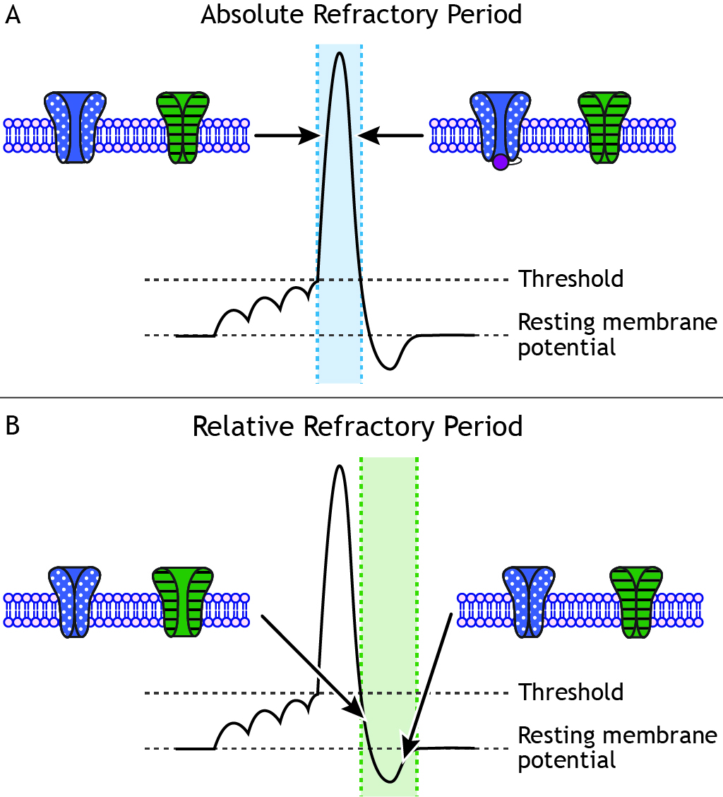
Each neuron does have a maximum firing rate. And even if the stimulus continues to increase in strength, the neuron cannot burn down at a college frequency. The maximum firing charge per unit of a jail cell is adamant by the status of the ion channels in the neuronal membrane during the different phases of the action potential. During the absolute refractory period, a second action potential cannot be fired under whatsoever circumstances regardless of the strength of the stimulus. The voltage-gated sodium channels are either open (during the rising phase) or inactivated (during the falling phase).
The Relative Refractory Period
When the cell repolarizes and the voltage-gated sodium channels de-inactivate and return to a closed state, the cell is once again able to fire another action potential. Even so, during the end of the falling stage and the during the undershoot, voltage-gated potassium channels are even so open. During the undershot, while the neuron is hyperpolarized, a larger-than-normal stimulus is needed to make the cell reach threshold once more. This segment of the action potential is chosen the relative refractory menstruation. Action potentials tin can exist fired, simply a stronger stimulus is needed than when the cell is at rest.
Activity Potential Characteristics
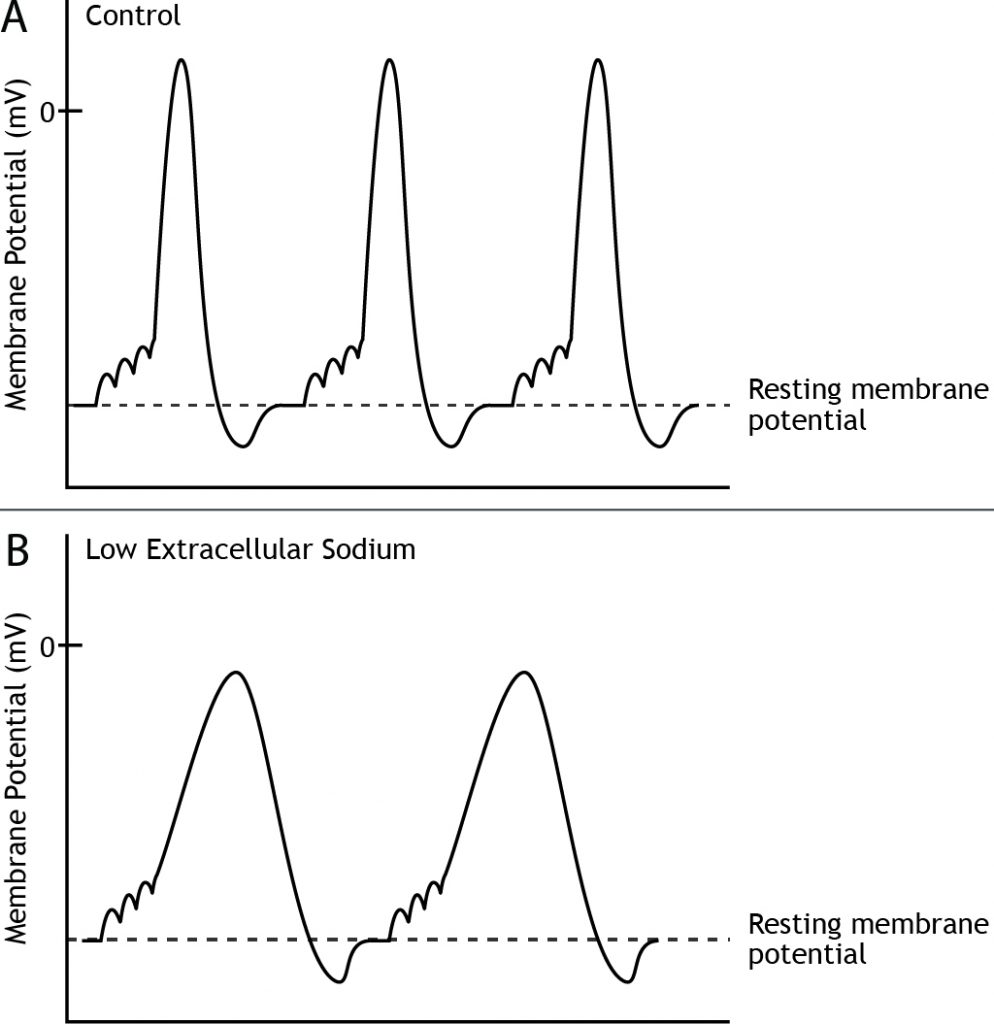
For a given prison cell, all action potentials accept the same characteristics; they depolarize to the same membrane potential value and accept the same amount of fourth dimension. However, different neurons may exhibit different action potential characteristics. Likewise, if a neuron has a change in its environment, like altered extracellular ion concentrations, the shape of the action potential would change due to a change in the electrochemical gradients. For instance, if the external concentration of sodium is decreased, the equilibrium potential of sodium, as well as the force of the electrochemical gradients volition change, which will result in a slower rate of ascent and a lower amplitude of the activeness potential.
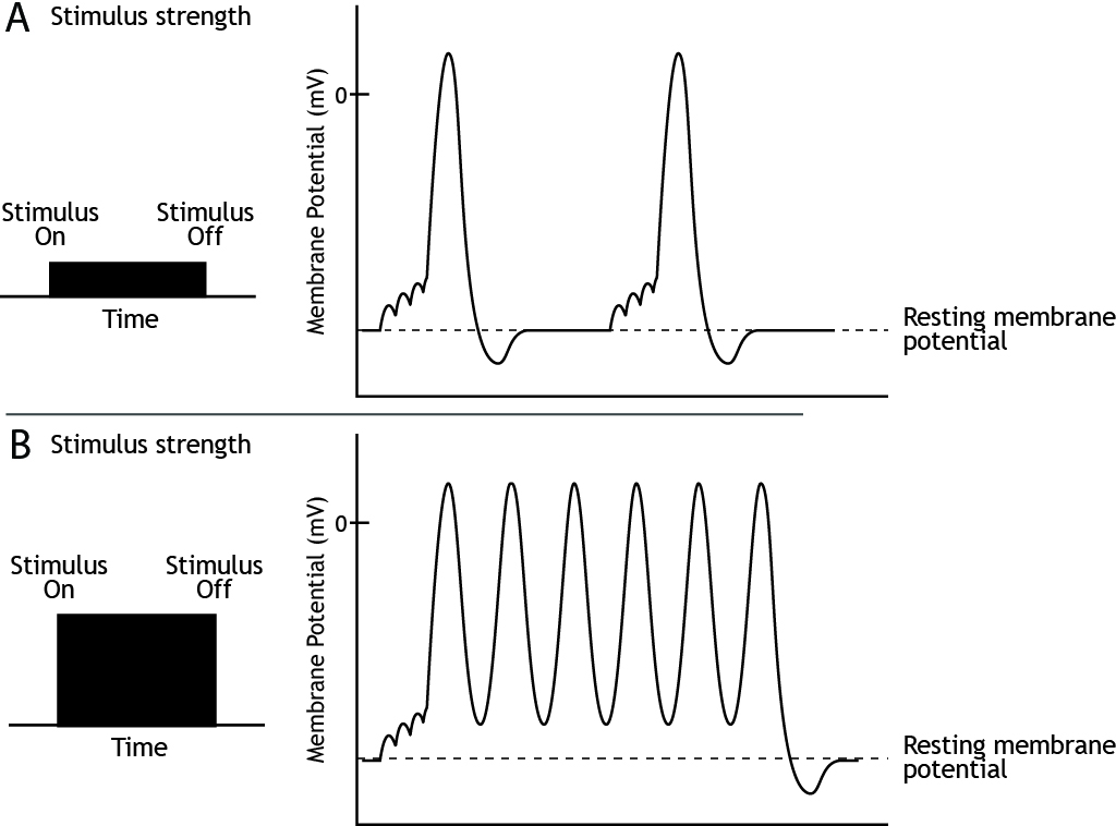
Stimulus Strength
The strength of a stimulus needs to be encoded by the neurons. We demand to exist able to perceive the difference, for example, between a dim light and a brilliant one. The frequency or rate of activity potential firing informs the nervous system of stimulus strength.
Since the summit of the action potential is ever the aforementioned for a given neuron, the strength of the stimulus is determined by the frequency of action potential firing. A weak stimulus would cause fewer action potentials to exist fired than a strong stimulus.
Management of Propagation
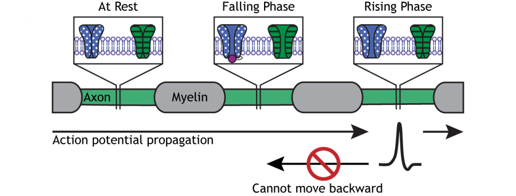
The action potential moves downwards the axon due to the influx of sodium depolarizing nearby segments of axon to threshold.
Blitheness 6.seven. A voltage change that reaches threshold volition cause voltage-gated sodium channels to open in the axonal membrane. The influx of sodium causes the rising phase of the activity potential, just the ion period also depolarizes nearby axon regions. As the depolarization reaches threshold, the action potential moves downwards the axon. The dotted, blue channels correspond voltage-gated sodium channels; the striped, dark-green channels represent voltage-gated potassium channels. 'Action Potential Motion' past Casey Henley is licensed under a Creative Commons Attribution Non-Commercial Share-Alike (CC-By-NC-SA) 4.0 International License. View static image of animation.
Activity potentials only motility in ane direction, though, from the cell body to the presynaptic terminal. The refractory menstruum keeps the activeness potential from moving astern down the axon. As the action potential moves from one Node of Ranvier to the side by side, the inactivated sodium channels in the previous axon segment forestall the membrane from depolarizing again. Therefore, the action potential can but move forward toward axon segments with closed sodium channels ready for ascent stage depolarization.
Speed of Propagation
Presence of Myelin
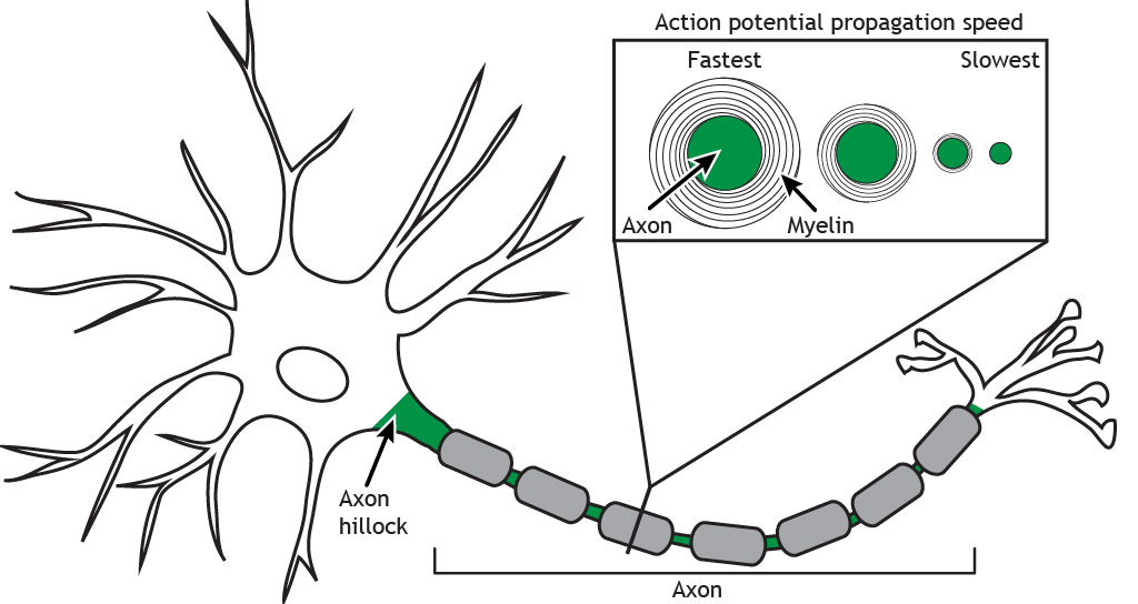
The presence of myelin leads to a significant increase in activity potential conduction speed compared to an unmyelinated axon. For a myelinated axon, the activity potential "jumps" between Nodes of Ranvier in a process chosen saltatory conduction. The nodes have a high density of voltage-gated channels, and the activity potential is able to skip the axon segments covered by the myelin. In an unmyelinated axon, the action potential moves in a continuous wave. In additional to the saltatory conduction process, the presence of myelin as well insulates the axon, preventing charge loss across the membrane, which also increases speed of the action potential.
Animation 6.eight. The action potential moves downwards an unmyelinated axon like a wave, opening voltage-gated channels forth the length of the axon. In a myelinated axon, though, the action potential is able to skip portions of the axon that are covered by the myelin; the action potential jumps from node to node and travels further down the axon in the same amount of fourth dimension. The dotted, blue channels stand for voltage-gated sodium channels; the striped, green channels stand for voltage-gated potassium channels. 'Action Potential Speed' by Casey Henley is licensed under a Creative Commons Attribution Not-Commercial Share-Akin (CC-BY-NC-SA) 4.0 International License. View static prototype of blitheness.
Diameter of Axon
The bore of the axon also affects speed. The larger the diameter of the axon, the faster the propagation of the activeness potential downward the axon. A larger axon leads to less resistance against the flow of ions, then the sodium ions are able to motility more apace to cause the regeneration of the action potential in the next axon segment.
- The voltage-gated ion channels are located along the axon hillock and axon; they open in response to the membrane potential reaching a threshold value
- The ascent phase of the action potential is a result of sodium influx
- The falling phase of the action potential is a result of potassium efflux
- Action potentials are all-or-none (postsynaptic potentials are graded)
- Action potential have the same height of depolarization for a given cell nether typical conditions
- The neuron cannot fire a second action potential during the absolute refractory phase
- The neuron tin can fire a second action potential during the relative refractory stage, but information technology requires a stronger stimulus than when the neuron is at rest
- Stimulus strength is coded by frequency of action potential firing
- Action potential travel in one direction due to the presence of inactivated voltage-gated sodium channels
- Speed of propagation relies on presence and thickness of myelin and bore of axon
Test Yourself!
Video Lecture
christensendaudgessed1982.blogspot.com
Source: https://openbooks.lib.msu.edu/neuroscience/chapter/action-potentials/
0 Response to "What Makes an Action Potential Rise to +35 Mv? What Makes It Drop Again After This Peak?"
Post a Comment